MyoQuant🔬: a tool to automatically quantify pathological features in muscle fiber histology images.

Project description


MyoQuant🔬: a tool to automatically quantify pathological features in muscle fiber histology images

MyoQuant🔬 is a command-line tool to automatically quantify pathological features in muscle fiber histology images.
It is built using CellPose, Stardist, custom neural-network models and image analysis techniques to automatically analyze myopathy histology images.
Currently MyoQuant is capable of quantifying centralization of nuclei in muscle fiber with HE staining, anomaly in the mitochondria distribution in muscle fibers with SDH staining and the number of type 1 muscle fiber vs type 2 muscle fiber with ATP staining.
An online demo with a web interface is available at https://huggingface.co/spaces/corentinm7/MyoQuant. This project is free and open-source under the AGPL license, feel free to fork and contribute to the development.
Warning: This tool is still in early phases and active development.
How to install
Installing from PyPi (Preferred)
MyoQuant package is officially available on PyPi (pip) repository. https://pypi.org/project/myoquant/
Using pip, you can simply install MyoQuant in a python environment with a simple: pip install myoquant
Installing from sources (Developers)
I recommend using UV for python environment management. See UV documentation.
- Clone this repository using
git clone https://github.com/lambda-science/MyoQuant.git - Create a virtual environment by using
uv sync - Run Myoquant with
uv run myoquant --help
How to Use
🆕 NEW: MyoQuant now comes with a Text User Interface (TUI) for an easier discoverability of the different CLI parameter. For this you can run myoquant tui or uv run myoquant tui to launch the TUI.

To use the command-line tool, first activate your venv in which MyoQuant is installed: source .venv/bin/activate or simply install the package using UV.
Then you can perform SDH or HE analysis. You can use the command myoquant --help or uv run myoquant --help to list available commands.
💡Full command documentation is available here: CLI Documentation
- For SDH Image Analysis the command is:
myoquant sdh-analysis IMAGE_PATH
Don't forget to runmyoquant sdh-analysis --helpfor information about options. - For HE Image Analysis the command is:
myoquant he-analysis IMAGE_PATH
Don't forget to runmyoquant he-analysis --helpfor information about options. - For ATP Image Analysis the command is:
myoquant atp-analysis IMAGE_PATH
Don't forget to runmyoquant atp-analysis --helpfor information about options.
_If you're running into an issue such as myoquant: command not found please check if you activated your virtual environment with the package installed. And also you can try to run it with the full command: python -m myoquant sdh-analysis --help or uv run myoquant sdh-analysis --help
Contact
Creator and Maintainer: Corentin Meyer, PhD in Biomedical AI contact@cmeyer.fr
Citing MyoQuant🔬
[placeholder]
Examples
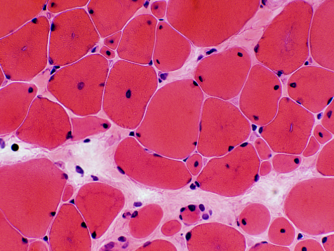
For HE Staining analysis, you can download this sample image: HERE
For SDH Staining analysis, you can download this sample image: HERE
For ATP Staining analysis, you can download this sample image: HERE
-
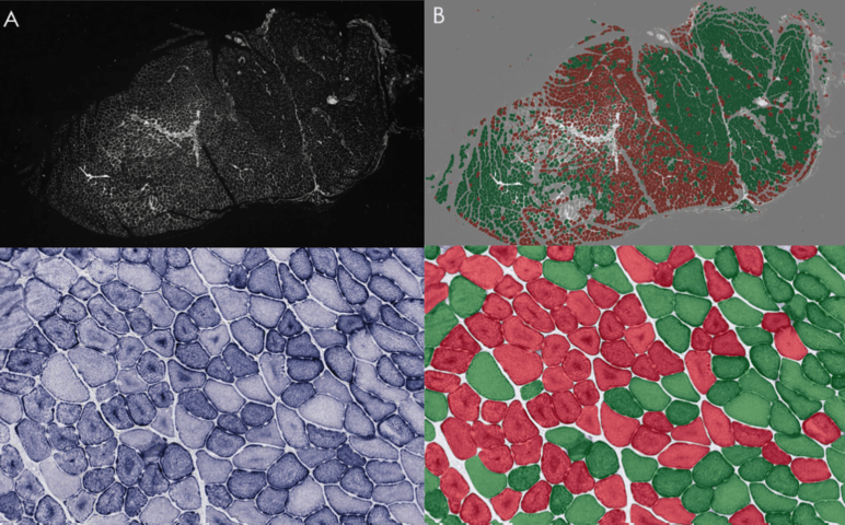
Example of successful SDH analysis output with:
myoquant sdh-analysis sample_sdh.jpg -
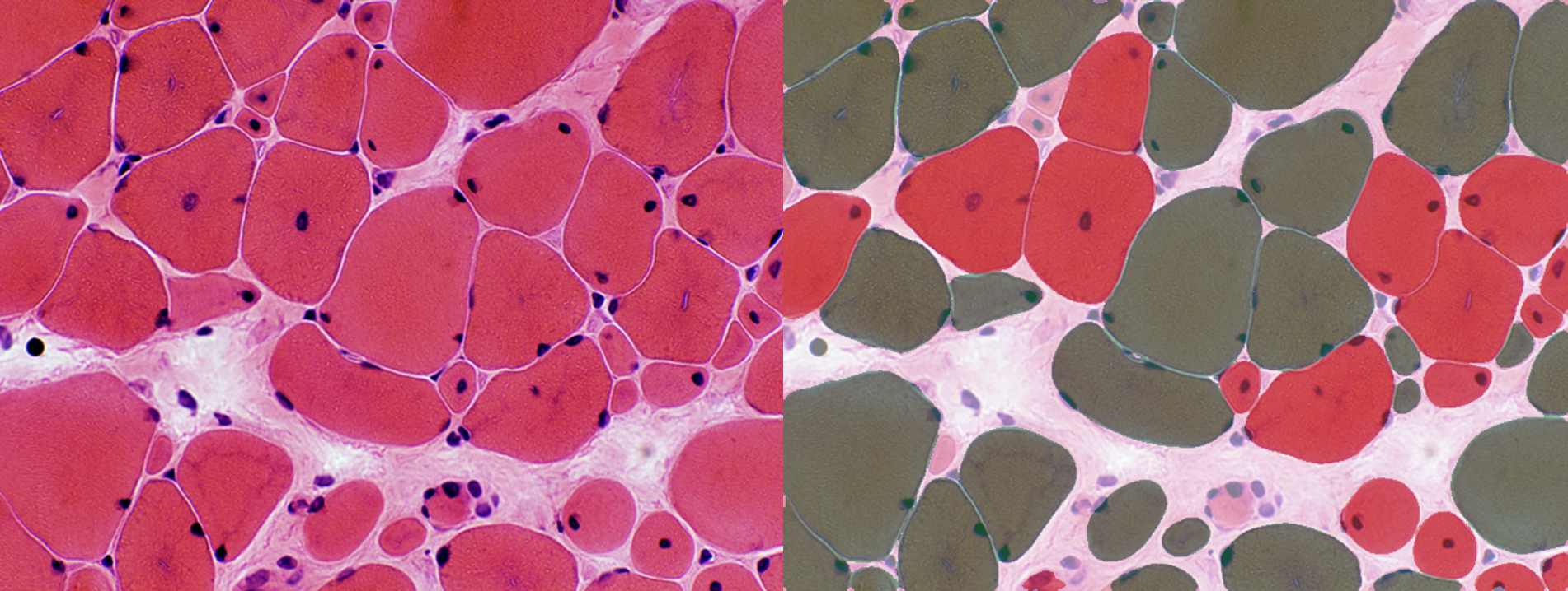
Example of HE analysis:
myoquant he-analysis sample_he.jpg

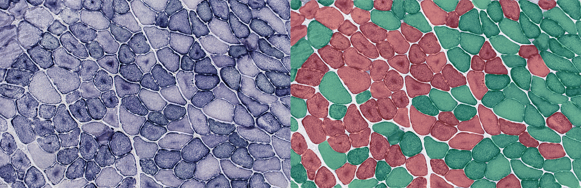
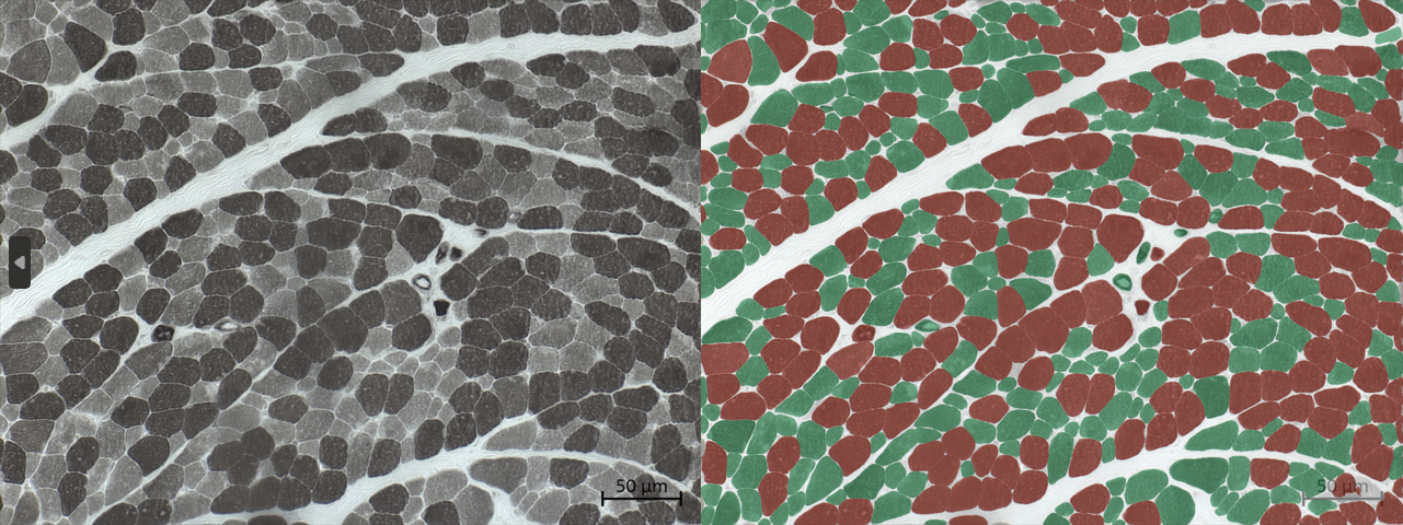
- Example of ATP analysis with:
myoquan atp-analysis sample_atp.jpg
Advanced information
HuggingFace🤗 repositories for Data and Model
In a effort to push for open-science, MyoQuant SDH dataset and model and availiable on HuggingFace🤗
Partners

MyoQuant is born within the collaboration between the CSTB Team @ ICube led by Julie D. Thompson, the Morphological Unit of the Institute of Myology of Paris led by Teresinha Evangelista, the imagery platform MyoImage of Center of Research in Myology led by Bruno Cadot, the photonic microscopy platform of the IGMBC led by Bertrand Vernay and the Pathophysiology of neuromuscular diseases team @ IGBMC led by Jocelyn Laporte
Release history Release notifications | RSS feed


Download files
Download the file for your platform. If you're not sure which to choose, learn more about installing packages.
Source Distribution
Built Distribution
Filter files by name, interpreter, ABI, and platform.
If you're not sure about the file name format, learn more about wheel file names.
Copy a direct link to the current filters
File details
Details for the file myoquant-0.4.0.tar.gz.
File metadata
- Download URL: myoquant-0.4.0.tar.gz
- Upload date:
- Size: 3.5 MB
- Tags: Source
- Uploaded using Trusted Publishing? No
- Uploaded via: uv/0.7.6
File hashes
| Algorithm | Hash digest | |
|---|---|---|
| SHA256 |
cd1e83af91a199e363ec305f1b70ceb2777368227d12697f82aafe0fde792823
|
|
| MD5 |
afdb79e092aec23740fd66fb5648fc7d
|
|
| BLAKE2b-256 |
c8ea33c8159d465f5538e93b0857c57bc4a9c3036caee7b4bab55c0e8e18715f
|
File details
Details for the file myoquant-0.4.0-py3-none-any.whl.
File metadata
- Download URL: myoquant-0.4.0-py3-none-any.whl
- Upload date:
- Size: 52.5 kB
- Tags: Python 3
- Uploaded using Trusted Publishing? No
- Uploaded via: uv/0.7.6
File hashes
| Algorithm | Hash digest | |
|---|---|---|
| SHA256 |
fbd0baf2a984dd2292204e5f6b9603123de68b84762844d98eec010c6c10eb1c
|
|
| MD5 |
67a5e899648b6ff3e83fc8dce0b689cf
|
|
| BLAKE2b-256 |
75d4ce4de0b692a107faf946b13ea45c99070718f2369e795cccdb3a35474b31
|


